| V.R. SYSTEMS Instrumentation Pvt. Ltd. |

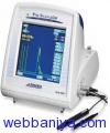
High Contrast Color Touch Screen • User Friendly Graphical Interface • 5 Programmable User Profiles • Scan Viewer Archiving Software • USB Interface • SD Memory Card • Built-In Thermal Printer • Storage Compartment • Portable Design Weighing 6 lbs (2.7kg) • Power Requirements: PacScan System: 100-240 VAC; 50/60 Hz Optional Printer: 100-240 VAC; 50/60 Hz 2. 300A+ A-Scan: Scan Modes: • Direct Contact/Immersion • 5 Examination Modes: Cataract Dense Cataract Aphakic Pseudophakic (5 settings) 4-Gate Manual Measurements: • ACD, Lens, Vitreous, Axial, AverageAxial, Standard Deviation • Individual Zone Velocities • Automatic Sensing Algorithm • Measure Review • Auto-Calibration Specifications: • Clinical Accuracy: ±0.10mm • Electrical Accuracy: ±0.023mm • Lens Calculations in 0.25D Increments • 10-Lens Database Formulas: • Refractive: Binkhorst Regression-II Theoretic/T Holladay Hoffer-Q Haigis • Post-Refractive: Latkany Myopic Latkany Hyperopic Aramberri Double-K A-Scan Probe Styles: • Standard A-Scan Probe for Hand-Held,Immersion, or Slit Lamp Mounted Application • Soft-Touch A-Scan Probe for Hand-Held Use Minimizing Corneal Compression
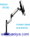
B-Scan Ultrasound Probes • Sealed 12 MHz or 20 MHz B-probes with focused transducers Scan Sampling • 256 ray scan Scan Controls • Fully adjustable time-varied gain (TVG), baseline, log gain, and exponential gain (e-gain) Scan Position Indicator • One-click selection of axial or longitudinal scan clock position with eye model confirmation • Free-form text for scan position details that automatically annotate onto images and video clips Video Clips • Capture and store 50-frame video clips at up to 20 frames per second (fps) • Replay in real-time, scalable slow motion, or one frame at a time • Store up to 6 video clips per eye per exam, easily add or remove video clips from exam record Images • Separately save any number of individual frames from video clips as images, complete with annotation(s) A-Scan Trace • Superimpose arbitrary A-scan trace onto images with a single button click Measurement • Unlimited measurements using linear calipers and angle measurement tool UBM Ultrasound Probe • HD water path probe with interchangeable focused transducers available in 35 or 50 MHz frequencies Scan Settings •Selectable scan setting profiles to optimize image quality for both sulcus-to-sulcus or angle detail Scan Sampling • 256 ray scan Scan Controls • Fully adjustable Scan Position Indicator • One-click selection of axial or longitudinal scan clock position with eye model confirmation • Free-form text for scan position details that automatically annotate onto images and video clips Video Clips • Capture and store 50-frame video clips at up to 20 frames per second (fps) • Replay in real-time, scalable slow motion, or one frame at a time • Store up to 6 video clips per eye per exam, easily add or remove video clips from exam record Images • Separately save any number of individual frames from video clips as images, complete with annotation(s) • Store an unlimited number of images for each eye per exam A-Scan Trace • Superimpose arbitrary A-scan trace onto images with a single button click and measure with calipers Measurement • Unlimited measurements using linear calipers and angle measurement tool A-Scan Ultrasound Probes • Sealed A-probe with 10 MHz focused transducer • Standard probe for immersion or soft-touch probe for direct contact with minimal corneal compression Scan Modes • Direct contact or immersion • Manual or Automatic Capture (Cataract, Dense Cataract, Aphakic, and Pseudophakic) Measurements • Anterior chamber depth (ACD), lens thickness, vitreous, and axial length (AXL) • Averages and standard deviation calculated for up to 10 scans per exam • Configurable zone tissue velocities IOL Formulas • Standard: Binkhorst, Regression-II, Theoretic/T, Holladay, Hoffer-Q, Haigis • Post-Refractive: Latkany Myopic Regression, Latkany Kyperopic, Aramberri Double-K
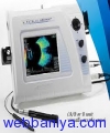
EZ5500+AB Scan 1. General: Display: • TFT Active Matrix Color LCD(262144 colors) • 6.5(17cm) Diagonal • 640 x 480 pixels • High Luminance (250:1) Video: • RS-170 BNC for video printer • VCR and remote viewing Size: 12.5W, 3.25H, 10.0D (31.7cm W, 8.2cm H, 25.4cm D) Weight: 5.25 lbs. (2.4kg) Voltage/HZ: 100/120/220/240 Volts and 50 Hz or 60 Hz auto sensed by input voltage Printer: High resolution video printer Date/Time: Built-in clock calendar Data Entry: Full alpha-numeric via touch screen 2. A-Scan: Probes: 10 MHz, focused, internal fixation light; Solid Tip or Soft-Touch Measurements: ACD, Lens, Vitreous, and Axial Length using individual zone velocities and moving gates Formulas: Holladay, Regression-II, Theoretic/ T, Binkhorst, Hoffer-Q, Haigis (optional) Modes: Automatic and Manual; Cataract, Dense Cataract, Aphakic & Pseudophakic (PMMA, Acryllic, Silicone, and Custom) Review: Stored A-Scan Patterns, A-Scan measurements, and statistics Statistics: Average, Std. Deviation, Range, and Maximum Difference from average Calculations: 6 constants per user profile, 9 user selected IOL powers vs. refraction, personalized A-constants and surgeon factors Displays: Multiple screens available for tabled, summarized and compared calculations Memory: Stores 5 scans and measurements, selected formula, IOL constants and user name Accuracy: • Electronic: ±0.023mm • Clinical ±0.1mm Range: • Automatic Mode: 18-33mm • Manual Mode: 0.5-35mm Calibration: Automatic with built-in calibration cylinder Report Data: Patient Name, ID #, Eye Examined, K-readings, User Name, Date, Time, Immersion On/Off Post Refractive Formulas: Latkany Myopic Regression, Latkany Hyperopic, Aramberri Double-K 3. B-Scan Probes: 10MHz, focused transducer, 30 frames/sec. Measurements: Distance and area Amplifier: 100 dB Gain, Logarithmic/ Linear/S-Curve, Gain, and TVG controls Magnification: Continuous Zoom (0.5x – 2.0x) with Pan (joystick controlled) Display Resolution: 640 x 480 pixels, color VGA with optimal tissue resolution of 0.15mm Processing: Reject below level, enhance contour and texture Freeze: Foot Pedal or touch screen activated Image: B-Scan with simultaneous selectable vector A-Scan Display: 60 Degree sector fan, 128 lines, Gray Scale, B/a presentation (B emphasized) or A/B (A emphasized), Gain TVG, Electronic Scale, Amplifier, OD/OS, Velocity, Probe Orientation, Patient and User Names, Date/Time • Maintains high resolution at all magnifications • Pan feature using built-in “joystick control • Gain and TVG controls for optimal diagnostic capability • Selectable Color or Gray Scale image • Software enhancement capability of frozen image • Selectable, simultaneous A-Scan vector • Sealed B-Scan probe provides smooth scanning with virtually no audible sound • 5 user selections 01
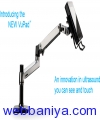
B-Scan Ultrasound Probes • Sealed 12 MHz or 20 MHz B-probes with focused transducers Scan Sampling • 256 ray scan Scan Controls • Fully adjustable time-varied gain (TVG), baseline, log gain, and exponential gain (e-gain) Scan Position Indicator • One-click selection of axial or longitudinal scan clock position with eye model confirmation • Free-form text for scan position details that automatically annotate onto images and video clips Video Clips • Capture and store 50-frame video clips at up to 20 frames per second (fps) • Replay in real-time, scalable slow motion, or one frame at a time • Store up to 6 video clips per eye per exam, easily add or remove video clips from exam record Images • Separately save any number of individual frames from video clips as images, complete with annotation(s) A-Scan Trace • Superimpose arbitrary A-scan trace onto images with a single button click Measurement • Unlimited measurements using linear calipers and angle measurement tool UBM Ultrasound Probe • HD water path probe with interchangeable focused transducers available in 35 or 50 MHz frequencies Scan Settings •Selectable scan setting profiles to optimize image quality for both sulcus-to-sulcus or angle detail Scan Sampling • 256 ray scan Scan Controls • Fully adjustable Scan Position Indicator • One-click selection of axial or longitudinal scan clock position with eye model confirmation • Free-form text for scan position details that automatically annotate onto images and video clips Video Clips • Capture and store 50-frame video clips at up to 20 frames per second (fps) • Replay in real-time, scalable slow motion, or one frame at a time • Store up to 6 video clips per eye per exam, easily add or remove video clips from exam record Images • Separately save any number of individual frames from video clips as images, complete with annotation(s) • Store an unlimited number of images for each eye per exam A-Scan Trace • Superimpose arbitrary A-scan trace onto images with a single button click and measure with calipers Measurement • Unlimited measurements using linear calipers and angle measurement tool A-Scan Ultrasound Probes • Sealed A-probe with 10 MHz focused transducer • Standard probe for immersion or soft-touch probe for direct contact with minimal corneal compression Scan Modes • Direct contact or immersion • Manual or Automatic Capture (Cataract, Dense Cataract, Aphakic, and Pseudophakic) Measurements • Anterior chamber depth (ACD), lens thickness, vitreous, and axial length (AXL) • Averages and standard deviation calculated for up to 10 scans per exam • Configurable zone tissue velocities IOL Formulas • Standard: Binkhorst, Regression-II, Theoretic/T, Holladay, Hoffer-Q, Haigis • Post-Refractive: Latkany Myopic Regression, Latkany Kyperopic, Aramberri Double-K Lens Selection • Lens calculations in 0.25D increments with built-in 1600+ lens database General Image Rendering • Outstanding B-Scan and UBM image quality using proprietary Enhanced Focus Rendering™ (EFR™) • Continuous interpolative zoom re-renders at each magnification level for optimized image quality (up to 4x zoom) Annotation • Automatic annotation of images and video clips Database • Full-scale patient database with exam record storage • Create and save individual user profiles with user-selectable defaults Reports • Detailed customizable exam reports for printing or exporting Hard Drive • 128 GB SSD solid-state drive Connectivity • 802.11n dual-band Wi-Fi and Bluetooth 4.0 • GigE Ethernet LAN and USB 3.0 ports • One touch export images (.jpg), video clips (.avi), and exam reports (.pdf) for referral, presentation, or EMR Printer • Any Windows-compatible printer Operating ASystem • Microsoft Windows 8 Console • Compact and streamline design with 10.1 high-resolution multi-touch monitor (1280 x 800 pixel) • Adjustable angle kickstand and VESA bracket for articulating arm or wall mounting Power • 100 - 240 VAC, 50/60 Hz auto-switching medical-grade power supply 1979 Marcus
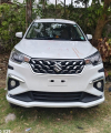
Book Mumbai Taxi Service for reliable taxi and cab services across Mumbai. Get local taxi, airport taxi, Mumbai airport

Mumbai Taxi Service provides convenient and dependable cab services across Mumbai for local travel, airport transfers, o
Mumbai Taxi Service provides convenient and dependable cab services across Mumbai for local travel, airport transfers, o
Mumbai Taxi Service provides convenient and dependable cab services across Mumbai for local travel, airport transfers, o
Mumbai Taxi Service provides convenient and dependable cab services across Mumbai for local travel, airport transfers, o
Mumbai Taxi Service provides convenient and dependable cab services across Mumbai for local travel, airport transfers, o

Book Mumbai Taxi Service for reliable taxi and cab services across Mumbai. Get local taxi, airport taxi, Mumbai airport

Srimathi Sundaravalli Memorial Arts and Science College (SSM), located in Perungalathur, Chennai, is committed to provid
Ayodhya Taxi Service provides reliable, comfortable, and convenient taxi services in Ayodhya for local travel, airport t

Does your parent or loved one need a disability assessment in Singapore for a CareShield Life, ElderShield or long-term
The Nicole Diamond Ring by Flawless Jewels New York combines timeless beauty with an elegant contemporary feel. This so
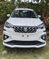
Banaras Taxi offers professional taxi service in Varanasi for tourists, families, local customers and business traveller

Banaras Taxi provides car rental and taxi services in Varanasi for local transportation, airport transfers, railway stat
Banaras Taxi is a reliable taxi service in Varanasi, providing comfortable and convenient transportation for local trave
Banaras Taxi is a reliable taxi service in Varanasi, providing comfortable and convenient transportation for local trave

Step Up Construction is a San Jose and Bay Area general contractor specializing in home remodeling, ADU construction, ki
MBBR MEDIA Modern wastewater treatment is moving toward compact and efficient biological systems, and mbbr media pla

Bhagyalaxmi Car Rental Hyderabad offers reliable, comfortable, and convenient car rental services for local travel, airp

car lockout service near me Car Lockout Service Lexington ky lockout service for car Lexington ky lockout service for
ABS Foods & Beverages is a Delhi-NCR based corporate food service company specializing in workplace dining, cafeteria ma